Dear all,
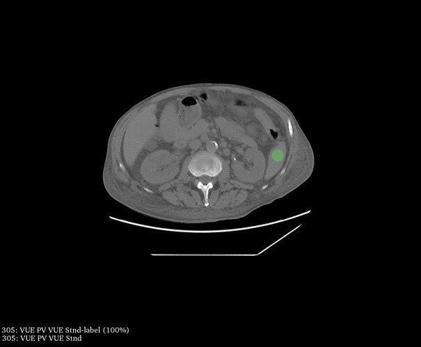
We are currently working on a project that involves obtaining pyradiomics features from dual-energy CT images which are in a normal DICOM format and can be loaded into 3DSlicer without any problems. We used the Pyradiomics 3DSlicer extension to compute results from 2D circular regions of interest (ROIs) in different organs. However, in each of the different kinds of images as well as in each of the ROIs we measured, we are getting ‘odd looking’ results for some of the examinations.
In the screenshot I attached, you see results obtained from an ROI in the lower spleen. Each row is one (different) CT examination. We are uncertain because there are examinations for which a lot of the features give the same numbers (often 0/1) without decimals – is this likely to be due to an issue with the extension and/or image or may these results actually be sound?
Thank you very much for your help - happy to provide additional information, if needed.
Simon


