I would be happy to have our R&D group responsible for the Artis software looking into this in detail.
Could you supply me with the original 3D data set / MRI volume?
As noted in the thread, the software copies attributed one to one from the original images into the Surface IOD. Therefore it is necessary to have the original data to troubleshoot efficiently.
Thank you very much.
For clarity, many programs with support for SINGLEBIT data (tried PixelMed, MircoDicom, Weasis, Aliza) can open that file standalone (setting spacing, origin, etc. to defaults).
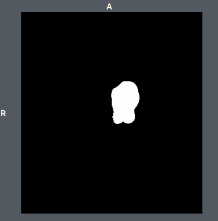
It looks like this (284x320x192), here slice #128 (PixelMed, the same in other programs)
As for the MR images, I am not able to provide the original images without further processing. The data comes from clinical collaborators working on a confidential brain imaging project, with the data not yet published. I do not have their permission to share the data. Another concern is that, although the metadata is already anonymized, with these MR head datasets, it is possible to reconstruct the facial features, which can violate the GDPR !
Would pydicom or another tool be able to zero out the pixel data and save the files without any changes to the DICOM metadata? If so, then I can send such cleaned out data. If you have experience with this on pydicom or another library, let me know. I could try it myself in a couple of days, but I have no experience which tool can clean up the pixel data while keeping the meta data untouched. Any ideas? Thanks.
@AshrafM I understand your valid concern. It would be great if you could dump the MR images as anonymized TEXT files. It should be sufficient for the analysis.
You can do this with mDicom (MicroDicom Viewer) for example. Under the file menu you can export a whole study or series as TEXT files. That would be one way of omitting pixel data.
Thank you for your suggestions. I wrote some code with pydicom to replace the pixel data with an ellipsoid, while trying to keep everything else in the dicom unchanged. I also extracted he DICOM meta data via both Slicer and mDICOM and I am providing the text files here.