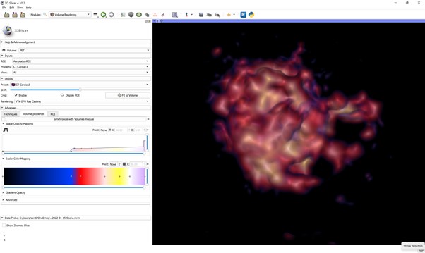
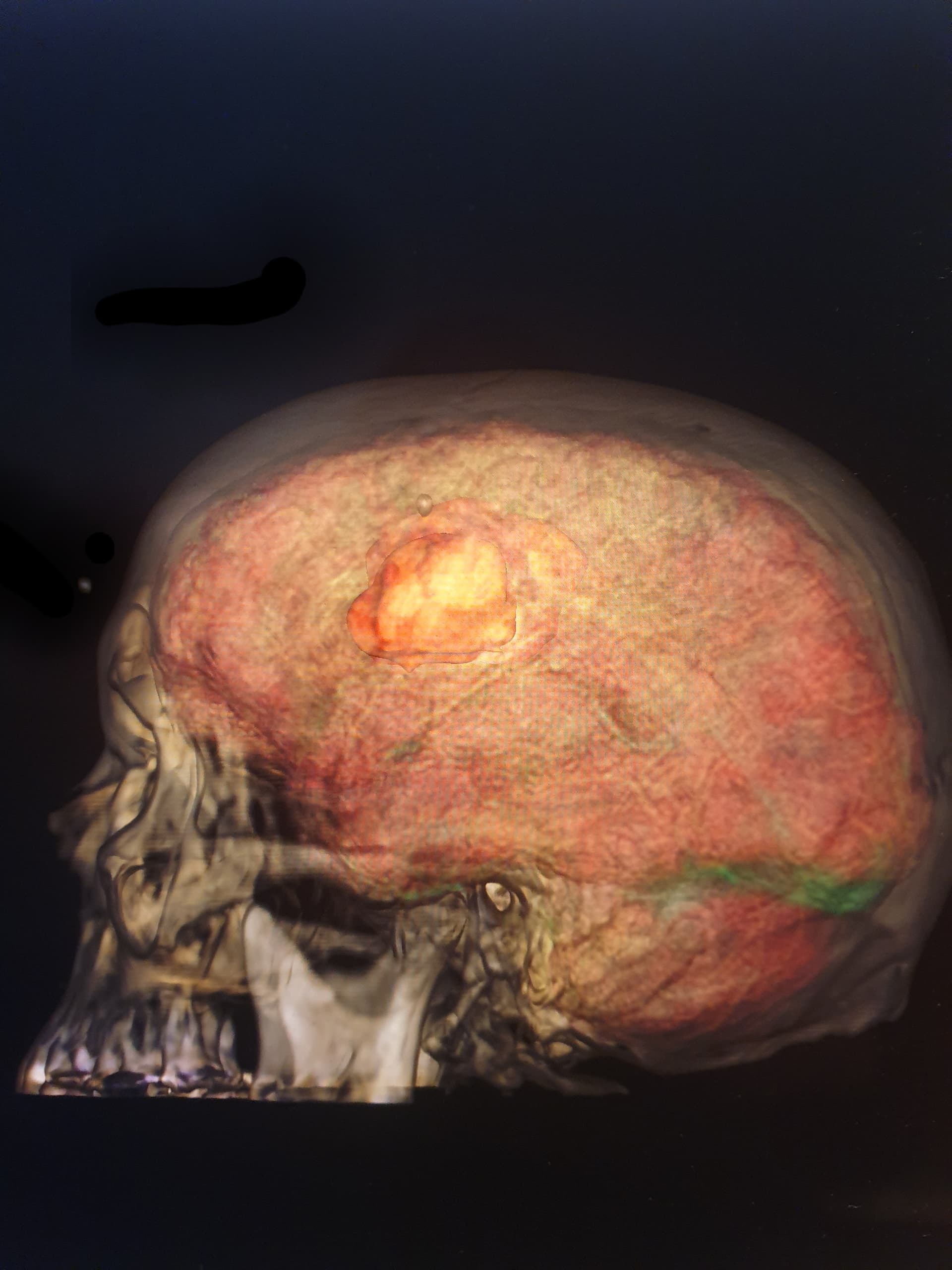
Just undertaken a project for my MSc in Medical Visualisation at Glasgow University. I had to register and render three datasets of a brain tumour using MRI, CT and PET. It was no easy feat given I am a complete newbie to Slicer and working on version 4.10. I was mainly guided by our Video lectures, which are great, but only give you a small amount of information, the rest is left for us to work & find out. So, I worked my way through many youtube/ online videos and managed to register the MRI with the Pet scan and then the MRI with the CT scan, with a lot of questions I needed to have answers to along the way to fulfil this project (I also during the three months have had other modalities and assignments to fulfil on 3dstudio max and unity and also Academic writing, so no mean feat by anyone standards to then fulfil 4 exercises in 3dslicer the brain being only one of them). So I worked my way through things and before I could render any of three datasets I had to get answers to my questions and the only way I could do that was to speak to a slicer expert! So I managed to get hold of Andras Lasso, who spent a few hrs of his time showing me better ways of doing things, and clarifying the answers to my dilemma which was how do we register the three data sets together, well we don’t was his simple answer, we only register two at a time!! Andras also explained that DVR is not tangible and that I need to load the TF to view it. So I was beginning to realise very quickly that knowing your way around the interface and understanding the lingo and what terminology means what is so important, but mostly it is trial and error. The MRI transformed data is not great to DVR, as it doesn’t show the brain surface or allow the viewer to visualise the spatial relationship, location and density of the tumour. I needed to have a better option than just the current MRI data set. So Andras introduced me to the Skull stripper you can find this in view/extension manager/skull stripper. It’s an amazing extension and really showcased the brain surface, I then added a pre-set, adapted the opacity and colour mapping to add depth and the gradient opacity to show the brain surface which also allowed a better understanding of the spatial relationship once I had adapted the CT and Pet Scans. So a massive shout out goes to Andras Lasso for all your amazing help and here are a few images from my project. I really love 3dSlicer and it can only get better from here, I love it so much that I have decided to use it for the thesis, no idea in what context yet, but I have time. Its been a steep learning curve with many challenges along the way, but learning from each and every one of them and having the amazing support from Andras has been unquantifiable. Enjoy the pics of the brain hop you can work out the tumour and see the metastasis and also the skull. Thank you, thank you, thank you Andras for your amazing support!!! Sandie


4 Likes