@kirezgik, @fedorov - so what is the problem? PK Modeling works for me with this data set.
What does not work is the “Output Fitted Data 4D” feature.
I did not realize this was the case. What AIF did you use?
Hi – sorry I was not aware that the frame time issue had been figured out (since it was not brought up when we discussed Andrew and Jihun’s mouse data).
Attached is the AIF label map that I used – voxels found using an automated algorithm applied to an artery.
Best
Sharon
Dear Dr Fedorov,
Hello again!
I am using pkModeling module on a liver tumor analysis project.
I have a question, that on Huang2014, it says pkModeling(BWH-3DSlicer) is using Tofts model. The paper was published 4 years ago.
I would like to make sure whether there is a upgraded model (for example Extended Tofts Model, or FXR-SSM) that is used in Slicer 4.9.0 nightly build version.
The question come up in my mind, because on this paper, it gives different definititions,
[Tofts model Huang14]=[Kety model Parker06]
[Extended Tofts model Huang14]=[Tofts model Parker06]
which confused me.
Thank you so much everytime!
–Tiger Hu
Tiger, we did not add any new models to PkModeling since the Huang2014 paper.
Dear Dr Fedorov,
Since there is an option of computing fpv(i.e vp) with 3-parameter model,

I assume you are using extended Tofts model, not Kety model

because extended Tofts model contains 3 parameters Ktrans, vp and ve, and Kety model only contains 2.
I will double check the results from calculation with pkmodeling module.
Thanks again!
-Tiger Hu
Both of those models (with and without fpv) were present at the time of the analysis reported in the Huang2014 paper, I think we just included one of those in the paper.
Dear Dr Fedorov,
Thank you for making it clear!
-Tiger Hu
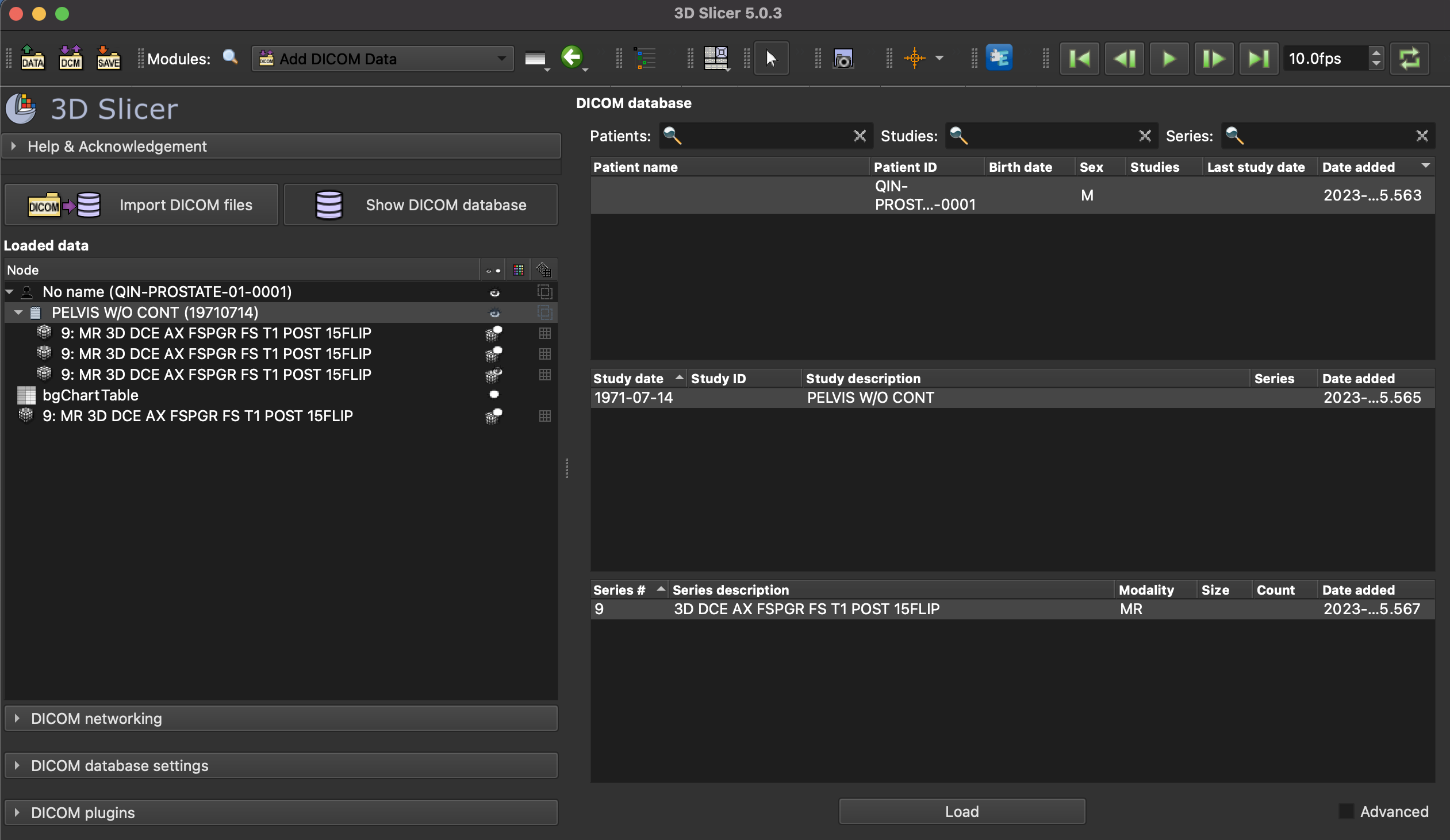
Actual behavior: Is there a way to do the quantitative analysis of DCE MRI of pancreatic tumor? I can only do semiquantitative analysis and create the plotting chart which shows the time-signal intensity curves of different ROIs, as it is discussed in this tutorial:
Yes, you can use the PkModeling extension for pharmacokinetic analysis of DCE

How can I extract the AIF mask, please